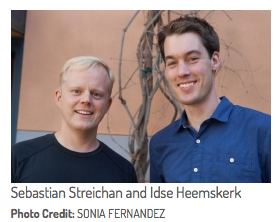
Two postdoctoral scholars from UCSB’s Kavli Institute for Theoretical Physics have developed a way to reduce dynamic bio-image data to two dimensions.
 (Santa Barbara, Calif.) — Today’s state-of-the-art optical microscopes produce voluminous three-dimensional data sets that are difficult to analyze. Now, two postdoctoral scholars from UC Santa Barbara’s Kavli Institute for Theoretical Physics (KITP) have developed a means of reducing data size and processing by orders of magnitude.
Taking advantage of the layered structure of many biological specimens, Sebastian Streichan and Idse Heemskerk created the Image Surface Analysis Environment (ImSAnE), a method that constructs an atlas of two-dimensional maps for dynamic tissue surfaces such as the early fruit fly embryo. Their findings appear today in the journal Nature Methods.
By implementing ImSAnE as an open source MATLAB toolbox, the KITP researchers provide a practical, highly accessible tool for data reduction and analysis of layered tissues. MATLAB is a high-level language and interactive environment used by millions of engineers and scientists to explore and visualize ideas and to collaborate across disciplines.
“We can now record the entire development of fruit flies from a couple hundred cells until a maggot hatches and crawls away — at a resolution that is good enough to track individual cells,” Streichan said. “Such data allows us to answer basic questions about developmental biology and the role of physics in shaping the developing body,” Heemskerk added.
