2025 Course Description

Course Subject:
As animals move their bodies to explore the world, they must integrate sensory and motor information. Course participants will conduct experiments in different invertebrate organisms and sensory systems to map the neuronal circuits and algorithmic principles underlying the sensory-motor control of active sensing. Research projects will include the design of new behavioral assays, behavior quantification, manipulation and measurement of neural activity, and advanced data analysis to develop physical models of behavioral control.
Course Directors:
Instructors:
Guest Lecturers (partial list):
The summer course is closely linked to the concurrent KITP program Neurophysics of Active Sensing. Course participants will attend the program's daily research seminars as part of the course curriculum. Students and lecturers will also have frequent opportunities for less formal interactions. Confirmed program participants include Florian van Breugel (UNR), Lisa Fenk (MPI-BI), David Kleinfeld (UCSD), Leo Petreanu (Champalimaud), Kathy Nagel (NYU), Pavan Ramdya (EPFL), Dima Rinberg (NYU), Monika Scholz (MPI-CAESAR), Tanya Tabachnik (Zuckerman Inst.) and Massimo Vergassola (UCSD).
Course Structure
The structure for this five-week school follows the model established in the 2018 course on sensory navigation and 2022 course on locomotion. The course will be closely linked to the concurrent KITP program "Neurophysics of Active Sensing", with students attending program talks each morning, as well as joint social events. Interaction with program speakers will also be facilitated through tutorial sessions and informal meetings over coffee and meals. Afternoon are dedicated to lab work with ample time for data collection and analysis. Teams of 4 students will work with 2 instructors and a teaching assistant on a research project for the full 5 weeks of the school. Students will also have room to explore their own interests, based on initial instructor-guided projects. Students in different modules will interact informally as many research areas benefit from comparison or use similar technical approaches. The philosophy of the school fosters knowledge sharing between students and instructors. Students actively teach and learn from each other, drawing on their diverse areas of expertise. Project groups are intentionally composed of students from different academic backgrounds, encouraging cross-pollination of ideas. Additionally, students work closely with instructors, teaching assistants, and guest lecturers from the associated KITP program, leveraging their research experience. At the end of the course, the outcome of the research projects will be presented to the KITP program for feedback.
A typical day's schedule includes:
- 2 morning program talks at KITP
- Lunch with other course and program participants
- Afternoon lab work
- Dinner in a dining hall, followed by evening lab work or an informal lecture or science discussion.
On weekends, students can attend program BBQs and self-organize outings to the beach or around Santa Barbara.
Accommodations, Fees and Financial Assistance
Students are housed in double-occupancy rooms in UCSB student apartments and provided with 19-meal plan at campus dining commons. The room and board fee is $2861. There are no other fees or tuition. Students who need financial assistance can request it in their course application form; financial need does not affect an applicant's chances of admission. A limited number of single rooms are available for a $1462 supplement.
Experimental Projects
A neural substrate for efference copy
Co-Instructors: Brad Dickerson* and Eugenia Chiappe
*module leader 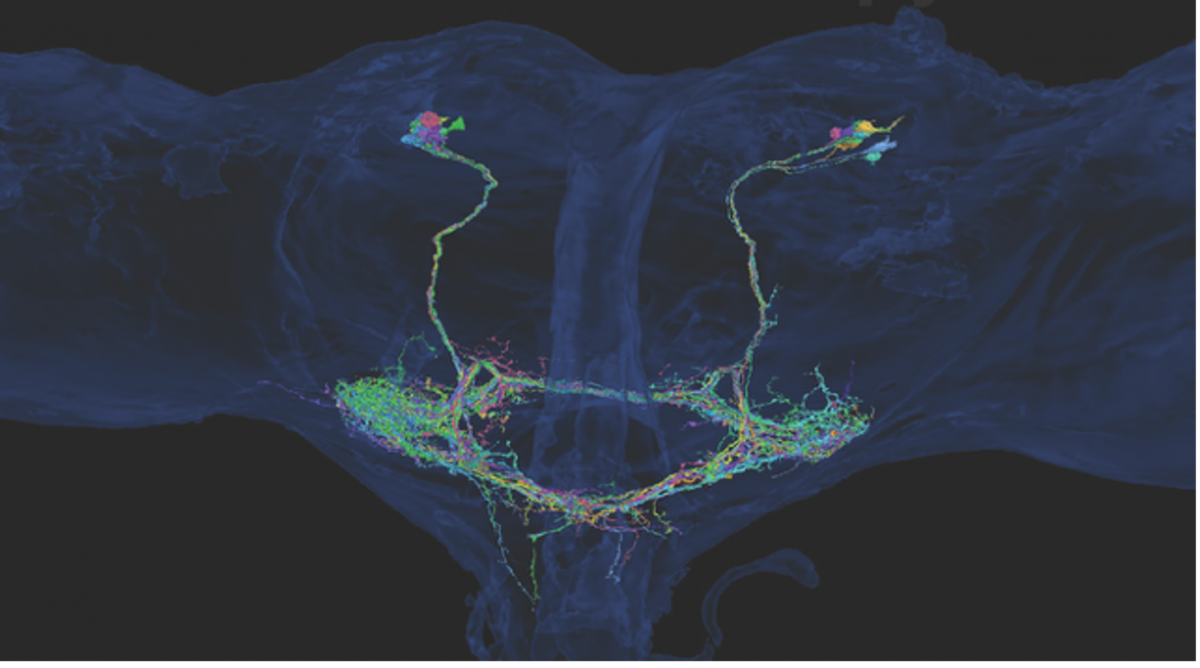
This module aims to uncover the neural mechanisms underlying the integration of self-generated motion and external sensory cues during voluntary sensorimotor behavior in flies. Previous work has shown that the fly visual system utilizes an "efference copy" signal to briefly attenuate the responses of motion-sensitive neurons during active flight maneuvers, but the origin of this signal remains unclear. We hypothesize that the fly's halteres, specialized structures that provide crucial feedback for flight control, transmit inhibitory signals to visual interneurons to enable the distinction between self-motion and external perturbations. By combining recent connectome data and genetic tools, we have identified a population of neurons, termed cLPTCrns, that provide this inhibitory input from the halteres to motion-sensitive neurons like the lobula plate tangential cells and ocelli. The goal of this module is to examine how manipulating the activity of these neurons affects the flies' ability to execute rapid, voluntary turning maneuvers (saccades) in the context of self-generated optic flow.
Understanding the functions of the descending information from mechanosensors in antennae to the haltere motor system
Co-Instructors: Jessica Fox*, Sung Soo Kim and Marie Suver
*module leader 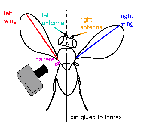
This module aims to elucidate the neural mechanisms underlying the control of haltere motion in flies. Previous work has shown that descending interneurons (DNs) in the central brain can modulate haltere kinematics, thereby indirectly regulating the mechanosensory feedback that drives wing and head movements
during flight. The research plan involves two key objectives: 1) selectively activating or inactivating DN populations, particularly the DNp17 neurons that project exclusively to the haltere neuropil, while observing resulting changes in haltere, head, and wing movements using high-speed videography; and 2) directly measuring the responses of these DNs to antennal airflow stimulation during tethered flight, using two-photon calcium imaging. These experiments are designed to engage students in a wide range of neuroscience research techniques, including anatomy, behavior, physiology, optogenetics, and data analysis, ultimately mapping the neural circuits linking brain activity to the control of flight-related behaviors in insects.
Evolution of the neuromechanics of active sensing in the Drosophila larva
Co-Instructors: Ellie Heckscher*, Matthieu Louis, Akinao Nose, Nikolaos Papachatzis and Madhu Venkadesan
*module leader 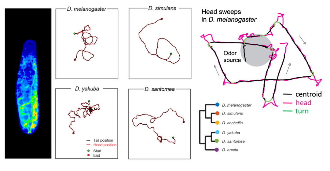
For survival, larval fruit flies must successfully navigate through their environments—locating nutritive food sources and avoiding biotic (e.g., predators) and abiotic (e.g., heat) threats using active sensing behaviors. One of the best understood of these behaviors in Drosophila melanogaster larvae is a head sweep (or head cast) that moves sensory organs laterally to scan the environment. Active sensing mechanisms rely on the larva’s ability to accurately monitor its body position in space (i.e., proprioception). Yet, how active sensing interacts with organismal biophysics and self-sensing remains elusive. We will approach this question by comparing the active sensing behaviors across different species of Drosophila and other related insect species and through mathematical analyses of self-motion-induced flow and elastic deformation of the body that affects proprioception. We will consider orientation responses to two sensory modalities: chemotaxis and phototaxis. In particular, we will examine whether and how the evolution of sensory systems and proprioception have contributed to active sensing to adapt to the properties of distinct ecological niches.
Figure: Drosophila larva (left). Examples of larval trajectories of four Drosophila species on an agarose substrate with the same stiffness (Elie Fink, Louis lab). Illustration of head sweep in D. melanogaster (right) from Gomez-Marin et al., Nat. Com. 2011.
Navigating in the Extremes: Exploring Sensory-Driven Taxis and Cryptobiosis Recovery in Tardigrades
Co-instructors: Molly Kirk and Ana Lyons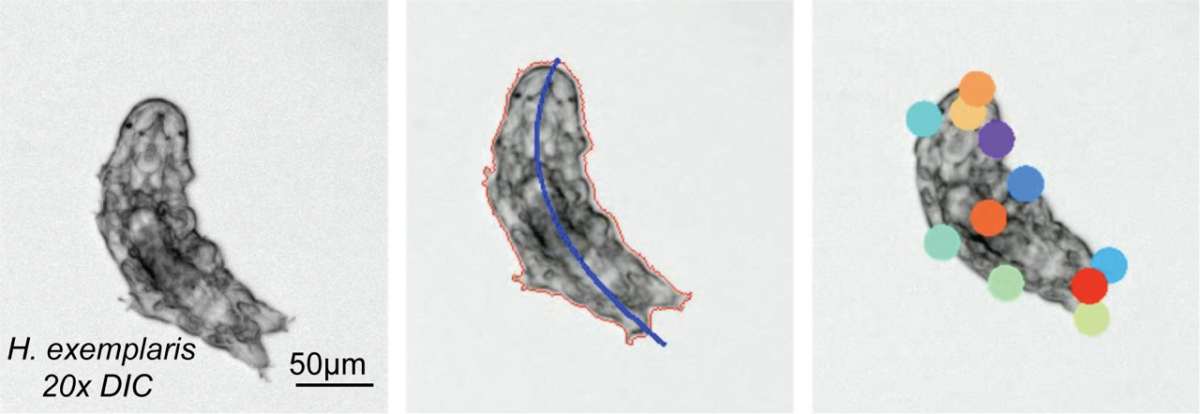
In this module, we will explore sensory-driven navigation (taxis) in tardigrade, Hypsibius exemplaris. Tardigrades are extraordinary micro-animals capable of withstanding extreme environmental conditions, including near-complete anoxia, desiccation, freezing in the absence of cryoprotectants, and even the vacuum and ionizing radiation of space. With a compact nervous system of approximately 200 neurons, tardigrades represent a simple yet powerful model to study sensory responses in environments that would be lethal to most other organisms. This module will investigate how tardigrades respond to various stimuli—optical, chemical, thermal, etc.—by identifying their behavioral strategies for taxis. Ultimately, we will explore how these navigational strategies change during recovery from cryptobiosis, a dormant state entered in response to extreme environmental stress, providing insights into the resilience of sensory systems in harsh conditions.
The module will focus on three unexplored areas of tardigrade neurobiology. (i) Valence Identification: By observing behaviors responding to various chemicals, light intensities, and temperature gradients, we will characterize the positive or negative associations that drive tardigrade movement. (ii) Behavioral Strategy: Through detailed behavioral tracking, students will learn to identify the distinct navigational strategies tardigrades employ when approaching or avoiding stimuli. (iii) Cryptobiosis Recovery Investigation: Lastly, the course will explore how sensory valences and navigational strategies are altered by cryptobiosis. Throughout the module, students will employ quantitative tracking techniques and analysis to understand how even simple nervous systems coordinate complex behaviors.
Antennal dynamics and active sensing in ants
Co-instructors: Venkatesh Murthy* and Orit Peleg
*module leader 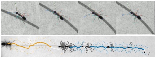
Ants, like many other insects, use their antennae actively to explore the world. Since they are largely terrestrial insects, they sample signals close to the ground – these signals are often in the form of chemicals. Trail following is characteristic behavior in many ants, and in this module, we will examine how ants follow trails deliberately constructed in the lab to allow precise quantification. Using videography, computer vision and computational ethology, students in this module will characterize trail following phenomena, focusing on antennal dynamics. Questions can center on the biology of trail following, empirical computational analysis to describe the behavior, or theoretical formulations to uncover algorithms used by ants to follow trails. In addition to behavior of solitary ants, students can also explore trail following by groups of ants to study collective behavior.
2024 Course Description

Application deadline March 24: Rolling review begins March 17
Course Subject:
Mobile genetic elements (MEs) can mediate rapid adaptation by introducing genes that encode novel functions related to pathogenesis, symbiosis and metabolism into host cells. MEs, and the accessory genes they carry, are as varied as the hosts and niches they are found within. Fuller understanding of the dynamics, constraints and consequences of ME movement will require interdisciplinary research merging computational and experimental approaches. This hands-on course will integrate laboratory experiment with quantitative bioinformatic analysis, providing students from diverse disciplinary backgrounds with new experience and skills that will assist them in developing rigorous, quantitative research in microbial evolution and ecology.
Course Faculty:
Joanne Emerson (UC Davis), Benjamin Good* (Stanford U.), Roberto Kolter (Harvard Medical School), Honour Mc Cann* (MPI-EB), Richard Neher (U. Basel ), Boris Shraiman (KITP/UCSB), and Paul Turner (Yale U.)
*Course Director
Guest Lecturers (partial list):
The summer course is closely linked to the concurrent KITP program Horizontal Gene Transfer and Mobile Elements in Microbial Ecology and Evolution. Course participants will attend the program's daily research seminars as part of the course curriculum. Students and lecturers will also have frequent opportunities for less formal interactions. Confirmed program participants include Aude Bernheim (Pasteur Inst.), Devaki Bhaya (Carnegie Science/Stanford U.), Allison Carey (U. Utah), Ilana Brito (Cornell U.), Daniel Fisher (Stanford U.) Nandita Garud (Stanford U.), Graham Hatfull (U. Pittsburgh), Daniel Huson (U. Tübingen), Eugene Koonin (NCBI), Michael Laub (MIT), Katie Pollard (Gladstone Inst., UCSF), Martin Polz (U. Vienna), Paul Rainey (MPI-EB), Eduardo Rocha (Inst. Pasteur), Julia Salzman (Stanford U.), and Erik van Nimwegen (U. Basel).
Course Structure
The course is 5 weeks long, and runs concurrently with the KITP Program Horizontal Gene Transfer and Mobile Elements in Microbial Ecology and Evolution. Students will begin with a bootcamp on basic wetlab techniques, then break into project groups for the first section of wetlab projects. After wrapping up wetlab data collection and preparing samples for sequencing, students break for a drylab bootcamp. Students will then join and complete a drylab project. During this period, sequencing for wetlab projects will be completed. During the last week, students will analyse the data from their wetlab projects, and present their findings to the program.
The experimental projects are led by instructors and TAs working closely with groups of 4-5 students. Additional guest lectures and training modules will be provided throughout the course. Students regularly attend morning lectures offered as part of the KITP Program, then head off for lab work in the afternoon and evening. Students are welcome to join visiting lecturers and program participants for tea breaks and evening BBQs at the Munger Physics Residence. Weekends are open for students to explore and enjoy the many recreational opportunities in the Santa Barbara area.
Accommodations, Fees, and Financial Assistance
There is no tuition, lab fee, or room and board fee. Admitted students are provided with double-occupancy rooms in UCSB student apartments and a 19-meal plan at campus dining commons. A limited number of single rooms are available for a $2064 supplement. Students who need financial assistance with travel expenses can request it in the application form; financial need does not affect an applicant's chances of admission.
Experimental Projects
Evolution of bacterial genomic structure (Richard Neher and Marco Molari)
 Bacteria exchange genetic material horizontally, duplicate DNA, delete parts or invert stretches of their genome resulting in complex patterns of genetic diversity. Despite the overall diversity and structural complexity, the order of essential genes in bacterial genomes tends to be highly conserved and most of the horizontally acquired diversity is concentrated in hot-spots. In this project, we will construct genome graphs of groups of recently diverged bacteria (specific sequence types) using PanGraph. PanGraph is a tool to detect shared homologous sequences between a large number of genomes and summarize it in a concise graph structure. Using this graph, we will quantify the rates at which horizontal processes add or exchange genes and will detect aforementioned hot-spots and characterize their sequence repertoire.
___________________________________________________________________________________________________________
Examining G x G x E interactions in evolution of bacterial-pathogen resistance to therapeutic phages (Paul Turner)
This project examines three lytic phages of Pseudomonas aeruginosa (PsA) that have been used successfully in FDA-approved emergency therapy to treat antibiotic-resistant pulmonary infections in humans. Each phage infects PsA using a different cellular receptor, and can select for host bacteria to evolve phage-resistance that either reduces virulence or switches hosts to become antibiotic sensitive (i.e., phage induced trade-offs). In week 1, students will challenge the phages to infect a collection of PsA strains that are cultured in different environments, to test how the culture conditions impact the spectrum of isolated phage-resistant mutants. These ‘rescue’ mutants will then be submitted for whole-genome sequencing, to analyze the loci underlying evolved phage-resistance. Week 2 will involve analyses of the genomic results. In addition, students will conduct phenotypic (growth) assays on the mutant isolates using automated spectrophotometers (microplate readers) and R programs that infer quantifiable bacterial traits, to conduct rigorous genotype-phenotype analyses. The overall goal is to generate a large dataset of novel results that will inform phage-bacteria-environment (G x G x E) interactions, useful for future emergency patient treatment leveraging phage-induced trade-offs in this important bacterial pathogen system.
___________________________________________________________________________________________________________
Dynamics of ICE movement in bacterial populations (Elena Colombi and Honour Mc Cann)
![]()
Integrative and conjugative elements (ICEs) are large, self-transmissible mobile elements that mediate the movement of ecologically significant genes across even distantly related membes of the Pseudomonas syringae species complex. Excision, conjugal transfer from the donor, and site-specific integration into the recipient can occur within fifteen minutes in vitro. Newly acquired ICEs can replace or recombine with ICEs already present in recipient cells, generating novel chimeric elements. ICEs are known to vector genes with functions associated with antibiotic resistance, pathogenicity and symbiosis, bacteriocin synthesis and more. For this project, we will start by performing transfer and competition experiments to assess the host range and fitness contributions of ICEs in different recipient strains. We will then set up and sequence a serial passage experiment to quantify rates of ICE transfer in mixed populations. Students will explore whether ICE proliferation is linked to their contributions to host fitness, or is selfish in nature.
___________________________________________________________________________________________________________
Rates of Horizontal Gene Transfer and Transposition in Non-Growing Bacteria (Roberto Kolter and Benedek Facskó)
![]()
Comparative genomics led to the insight that horizontal gene transfer events are the prevalent mode of bacterial genome evolution. We know much about the modes of horizontal gene transfer, i.e., transformation, conjugation, transduction, etc. Most of the past studies that measured the rates of these processes utilized fast growing populations. Yet, we know that in natural settings, bacteria seldom encounter conditions propitious for fast growth. In this project, we will quantitate the rates of transduction in non-growing or very slow-growing bacterial populations. As a complement, we will also follow the dynamics of mobile genetic elements in non-growing cells. In addition, we'll explore the ecological implications of the little-explored phenomenon of abortive transduction.
___________________________________________________________________________________________________________
Spatial patterns of soil viral communities (Joanne Emerson)
Students will investigate spatial structuring of soil viral communities to identify patterns and drivers of community composition. The project will begin with sampling soil and wetland environments in the area, then enriching for viral communities via filtration. Students will extract DNA from filtered and unfiltered samples, then sequence the viromes and metagenomes using MinION and Illumina platforms. Viral genomes will then be identified, and viral community composition will be compared to identify ecological differences along the sampled gradients. Finally, students will look for evidence of horizontal transfer by searching for integrated prophages in microbial genomes and 'host' genes in viromes.
2023 Course Description
Application deadline: February 26, 2023
Course Subject:
Animal morphogenesis, the question of how body parts acquire their form, remains a fascinating open problem of developmental biology and physics. Molecular analysis uncovered how the basic architecture of the body plan is laid out and how genetic patterning instructs cell fate. Quantitative investigation of cellular flow in vivo indicates that feedback mechanisms between genetics, mechanical force generation, and geometry ensure a robust and reproducible time course of shape. These analyses rely on studying embryogenesis in model organisms such as the fruit fly, mouse, or zebrafish. While superb genetic perturbations are readily available, physical analysis, such as measurements or even perturbation of mechanics, remains a high hurdle.
Cell biology successfully incorporated experimental methods from physics and engineering into the study of immortalized cell lines. Culturing techniques enable precise regulation and measurements of biochemical factors, as well as physical, and geometric constraints. Recent progress with stem cell culture now enables addressing fundamental questions of developmental biology, about the interplay of mechanics, biochemical patterning, and geometry. This course will study synthetic morphogenesis by integrating quantitative data analysis and mathematical modeling, in a variety of hands-on projects. Experimental systems include the mechanobiology of human embryonic stem cell patterning, mechanical pattern formation in the avian dermis, synthetic morphogenesis with mouse as well as human embryonic stem cell lines, and the regenerative model system hydra.
Course Directors:
Instructors:
Idse Heemskerk (Univ. of Michigan), Kinneret Keren (Technion), Alan Rodrigues (Rockefeller ), Amy Shyer (Rockefeller), and Vikas Trivedi (EMBL Barcelona)
Guest Lecturers (preliminary list):
The summer course is closely linked to the concurrent KITP program Dynamics of Self-Organization in Animal and Plant Development. Course participants will attend the program's daily research seminars as part of the course curriculum. Students and lecturers will also have frequent opportunities for less formal interactions. Confirmed program participants include Dominique Bergmann (Stanford), James Briscoe (Crick Inst.), Francis Corson (ENS Paris), Cassandra Extavour (Harvard), Ken Irvine (Rutgers), Thomas Lecuit (College de France/IBDM), Ani Kicheva (IST Austria), Prisca Liberali (FMI), Adam Martin (MIT), Elliot Meyerowitz (Caltech), Rashmi Priya (Crick Inst.), Sharad Ramanathan (Harvard), Jochen Rink (MPI-NAT), Adrienne Roeder (Cornell), Timothy Saunders (Warwick), Eric Siggia (Rockefeller), Katharina Sonnen (Hubrecht Inst.), David Sprinzak (Tel Aviv), and Aryeh Warmflash (Rice).
Course Structure
Students will attend Dynamics of Self-Organization in Animal and Plant Development program talks each morning and spend afternoons and evenings working in their lab groups and attending modeling and analysis tutorials. Interaction with program speakers will also be facilitated through tutorial sessions and informal meetings over coffee and meals, as well as social evenings held jointly with the program. Students will be matched to experimental modules based on their ranked choices and have ample room to explore their own interests, based on initial instructor-guided projects. Students in different modules will interact informally – many research areas benefit from comparison or use similar technical approaches. Students teach each other, sharing their different expertise--project groups will intentionally include students from different academic backgrounds--and work closely with expert instructors, TAs. and guest lecturers from the associated KITP program.
There is no course fee, and students are provided with housing at UCSB's Manzanita Village residence halls and a 19-meal plan at campus dining commons. A typical day's schedule includes:
- 2 morning talks at KITP
- Lunch in a dining hall or campus cafe, with other course or program participants
- Afternoon lab work
- Dinner in a dining hall, followed by evening lab work and/or science discussions
On weekends, students can attend program BBQs and self-organize outings to the beach or around Santa Barbara.
Experimental Projects (partial list)
Amy Shyer and Alan Rodrigues: Morphological symmetry breaking in self-organizing mesenchymal tissue
While much of the recent advances in our understanding of mechanics in development have focused on epithelial tissues, the Shyer-Rodrigues Laboratory of Morphogenesis studies morphological symmetry breaking driven by self-organizing mesenchymal tissues. Using a first-of-its-kind experimental platform, we combine live-cell imaging with collagen substrates specifically engineered to enable skin progenitor cells to self-organize into follicle patterns (https://www.quantamagazine.org/embryo-cells-set-patterns-for-growth-by-pushing-and-pulling-20220712/). By recapitulating a dynamic in vivo process, we will observe and manipulate multicellular activity in a tissue as it is generated.
___________________________________________________________________________________________________________
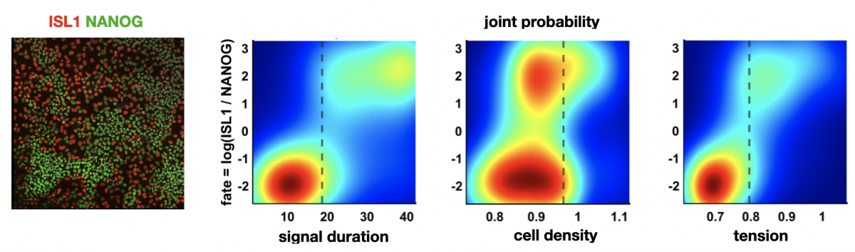
Figure: left: cells stained for fate markers after 42h of differentiation, right: (partially hypothetical) probability distributions showing how different factors relate to fate
Idse Heemskerk: Predicting cell fate in pluripotent stem cells
Pluripotent stem cells have remarkable capacity to self-organize into heterogeneous structures consisting of multiple cell types. Quantitative understanding of what makes one cell differentiate to one fate and a neighboring cell to a different fate is in most cases lacking. Variations in initial cell state, differences in morphogen signaling, and the mechanical environment all contribute, but their relative contributions and interdependence are often difficult to dissect.
In the context of the first cell fate decisions after pluripotency loss, associated with gastrulation, we will determine using single cell analysis of live and fixed imaging data how much redundant and non-redundant information different factors contain about cell fate. For example: does cell geometry at the onset of differentiation predict fate? What about the morphogen signaling dynamics? If so, does geometry predict morphogen signaling dynamics or are they independent inputs? And how do these relationships depend on substrate stiffness and intercellular forces?
Left figure: cells stained for fate markers after 42h of differentiation. Right figures: (partially hypothetical) probability distributions showing how different factors relate to fate. Figures by Seth Teague, unpublished.
___________________________________________________________________________________________________________
Kinneret Keren: Cytoskeletal dynamics in regenerating Hydra
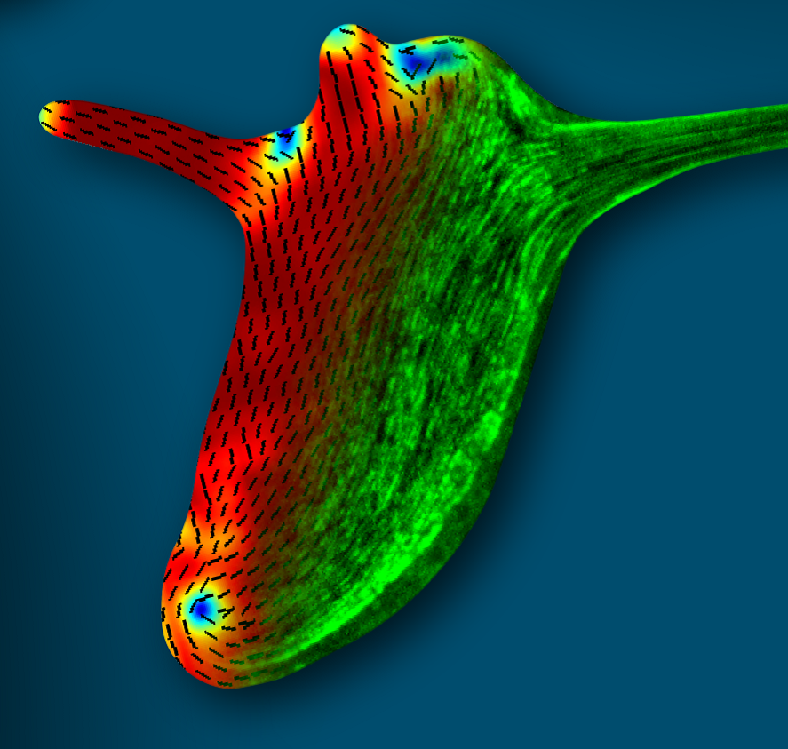
Figure: The supracellular actin fibers in a small regenerated Hydra (green) plotted together with the orientation field (black lines) and the nematic order parameter (jet colors).
Hydra is a small fresh-water animal renowned for its extraordinary whole-body regeneration capabilities. We will study the emergence of large-scale order in the cytoskeleton of regenerating Hydra, focusing on the establishment of parallel arrays of supracellular contractile actomyosin fibers. We will excise tissue pieces from transgenic Hydra expressing lifeact-GFP in the ectoderm and/or endoderm, and follow the dynamic organization of the actin cytoskeleton during regeneration using live microscopy. We are particularly interested in understanding how the large-scale nematic order develops in orthogonal directions in the two epithelial tissue layers, and how this relates to the morphogenesis process.
___________________________________________________________________________________________________________
Vikas Trivedi: Biophysics and metabolism in self-organising structures from embryonic stem cells
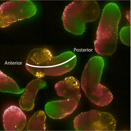 Figure: Hybridisation chain reaction (HCR) staining of 96h gastruloids. RNA expression patterns of Brachyury (green - primitive streak/mesodermal), Sox2 (red - pluripotent/ectodermal) and Sox17 (yellow - endodermal). The white line indicates the anterior-posterior axis illustrating the spatial organisation of cell fates in the gastruloid model.
How can tissue shapes and patterns emerge reproducibly and robustly in multicellular systems like animals? Despite more than 100 years of embryology, it still remains unclear how gene networks, forces and mechanical properties integrate with the metabolic state of the cells to self-organize complex structures. This is due to our inability to disentangle the combined action of these factors (biophysical properties, gene networks and metabolic activity) within populations of genetically equivalent cells. For example, the activity of genetic programs is accompanied by emergence of distinct biophysical characteristics within different cell types. At the same time the metabolic state of a cell can influence cell fate decisions by modifying transcription factor signalling, the epigenetic landscape as well as cellular mechanics. Making quantitative measurements and systematic perturbations of these factors are nearly impossible in vivo. Therefore, we will take advantage of aggregates of mouse embryonic stem cells that recapitulate hallmarks of early embryonic development in vitro. This will allow us to explore the role of metabolism in symmetry breaking, germ layer specification and emergence of distinct biophysical properties that mould the tissue. We will use machine-learning based image analysis, quantitative measurements of relative material properties and genetically engineered cells with live imaging to dissect how coordinated changes in metabolic states and biophysical properties affect morphogenesis.
___________________________________________________________________________________________________________
Idse Heemskerk, Sebastian Streichan and Xavi Trepat: Linking fate and force in micro patterned human pluripotent stem cells
Micropatterned colonies of human pluripotent stem cells (hPSCs) recapitulate aspects of human embryonic development. Such systems provide an opportunity to study developmental events that are otherwise inaccessible to quantitative experimental approaches. This cell culture environment offers new interrogation and perturbation approaches that promise insight into the dynamics early human morphogenesis. These include spatial force measurement maps, tuning substrate stiffness, and shape as well as size of the micropatterned hPSCs colonies. We seek to exploit these advantages and leverage synergies between groups to study the interplay of biochemical signaling, tissue mechanics, and geometry. Specifically, we will have a project studying how the interplay between secreted factors and mechanical forces drives the formation of primordial germ cells in humans. Another example will investigate how mechanical stresses are coordinated to achieve neural plate folding into a tube.